
![]()
![]()
![]()
![]()
![]()
![]()
![]()
![]()

![]()
ESI (electron spectroscopic imaging)&
EELS (electron energy loss spectroscopy)![]()
When passing through a specimen, electrons interact with it. Some are scattered without losing energy (elastic scattering), others lose energy inside the specimen by interacting with the electrons of the specimen (inelastic scattering). Several of the beam electrons lose exactly the amount of energy needed to eject a specific specimen electron. When the energy-loss electrons are registered according to their energy, they form an energy-loss spectrum, with a characteristic fall off in intensity, and superimposed elemental peaks (EELS). By using selected energy-loss electrons for imaging, elemental maps can be obtained (ESI).
The example shows fibers including calcium containing nano particles. The images on top are taken using energy-loss electrons in front and on the calcium elemental peak. For the fibers both images are nearly identical, but the particles appear much brighter if imaged using the energy-loss electrons of the calcium element peak. The difference between the images gives the elemental distribution which is shown in the lower right picture. The spectrum below shows the EELS spectrum with the calcium elemental peak.
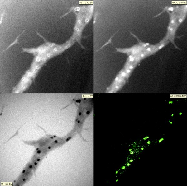
ESI for calcium
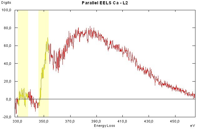
EELS
back to TEM gallery